Summary information and primary citation
- PDB-id
-
4wfm;
DSSR-derived features in text and
JSON formats
- Class
- RNA
- Method
- X-ray (3.1 Å)
- Summary
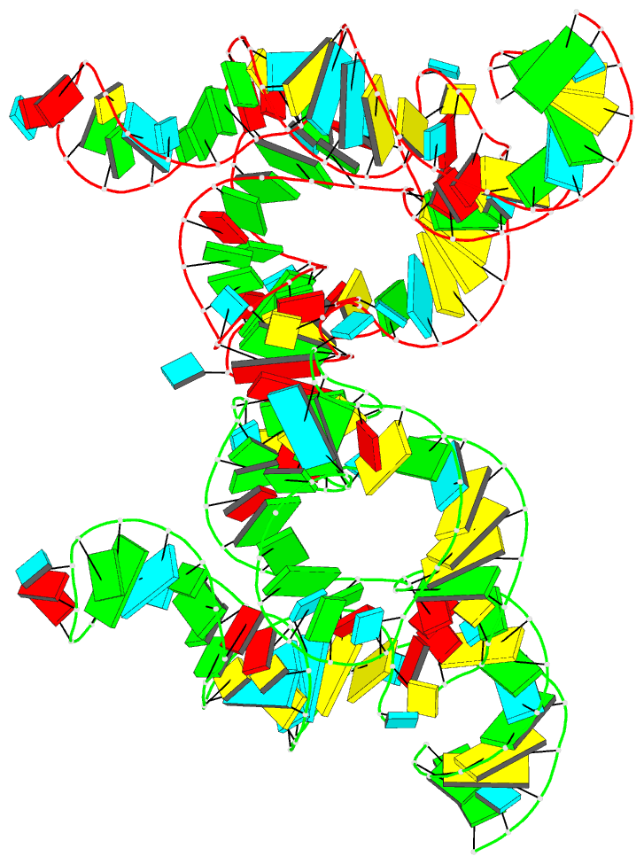
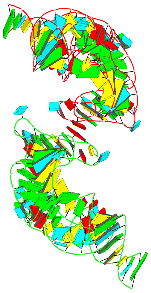
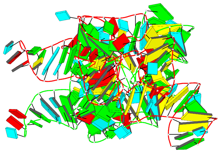
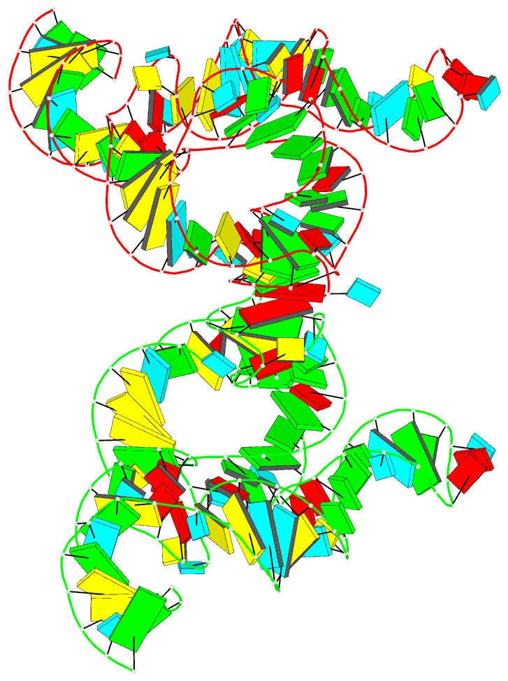
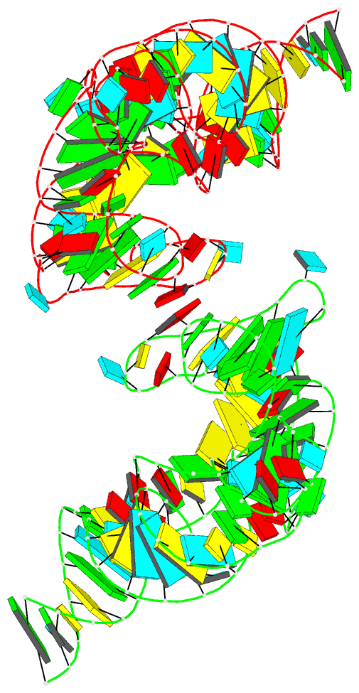
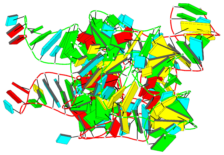
- Structure of the complete bacterial srp alu domain
- Reference
-
Kempf G, Wild K, Sinning I (2014): "Structure
of the complete bacterial SRP Alu domain."
Nucleic Acids Res., 42,
12284-12294. doi: 10.1093/nar/gku883.
- Abstract
- The Alu domain of the signal recognition particle (SRP)
arrests protein biosynthesis by competition with elongation
factor binding on the ribosome. The mammalian Alu domain is
a protein-RNA complex, while prokaryotic Alu domains are
protein-free with significant extensions of the RNA. Here
we report the crystal structure of the complete Alu domain
of Bacillus subtilis SRP RNA at 2.5 Å resolution. The
bacterial Alu RNA reveals a compact fold, which is
stabilized by prokaryote-specific extensions and
interactions. In this 'closed' conformation, the 5' and 3'
regions are clamped together by the additional helix 1, the
connecting 3-way junction and a novel minor groove
interaction, which we term the 'minor-saddle motif' (MSM).
The 5' region includes an extended loop-loop pseudoknot
made of five consecutive Watson-Crick base pairs. Homology
modeling with the human Alu domain in context of the
ribosome shows that an additional lobe in the pseudoknot
approaches the large subunit, while the absence of protein
results in the detachment from the small subunit. Our
findings provide the structural basis for purely RNA-driven
elongation arrest in prokaryotes, and give insights into
the structural adaption of SRP RNA during evolution.